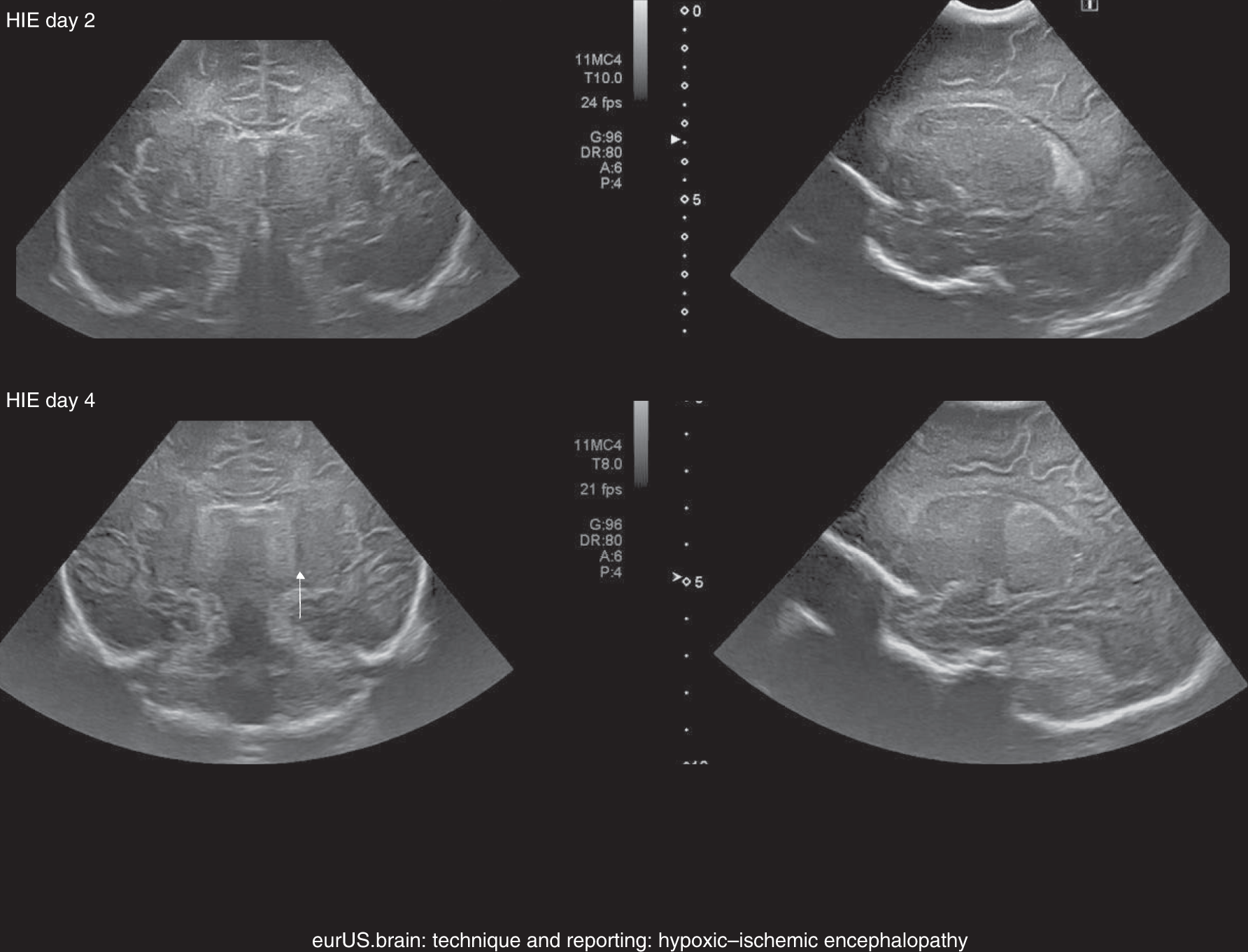
Home

Veramente esempio accuratamente coronal view ultrasound Decisione confront boccale

Ultrasound of acquired posterior fossa abnormalities in the newborn |  Pediatric Research
Ultrasound of acquired posterior fossa abnormalities in the newborn | Pediatric Research

14 Baby Brains ideas | hemorrhage, radiology, ultrasound sonography

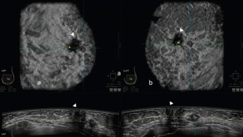
Automatic breast ultrasound: state of the art and future perspectives -  ecancer
Automatic breast ultrasound: state of the art and future perspectives - ecancer

The value of coronal view as a stand-alone assessment in women undergoing  automated breast ultrasound | SpringerLink
The value of coronal view as a stand-alone assessment in women undergoing automated breast ultrasound | SpringerLink

Ultrasound Machine Basics-Knobology, Probes, and Modes - POCUS 101

OB Images
OB Images

Scanning Planes and Scanning Methods | Radiology Key

ALiEM Cards
ALiEM Cards

Coronal view as a complementary ultrasound approach for prenatal diagnosis  of aberrant right subclavian artery - De León‐Luis - 2012 - Ultrasound in  Obstetrics & Gynecology - Wiley Online Library
Coronal view as a complementary ultrasound approach for prenatal diagnosis of aberrant right subclavian artery - De León‐Luis - 2012 - Ultrasound in Obstetrics & Gynecology - Wiley Online Library

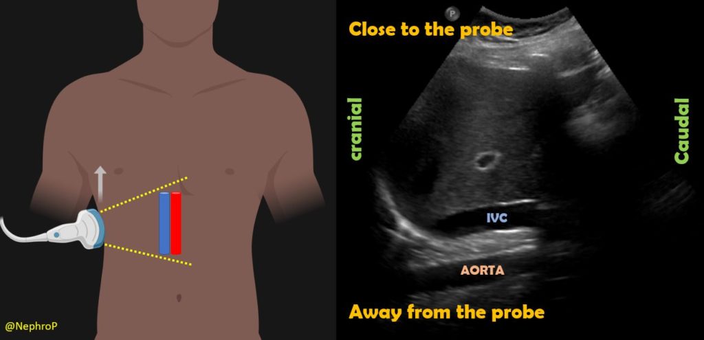
Inferior Vena Cava POCUS: The Basics of Image Acquisition - Renal Fellow  Network
Inferior Vena Cava POCUS: The Basics of Image Acquisition - Renal Fellow Network

Normal left kidney. Longitudinal coronal transducer position (A) and  long-axis ultrasound image of the kidney (B). Transverse coronal transducer  position. - ppt download
Normal left kidney. Longitudinal coronal transducer position (A) and long-axis ultrasound image of the kidney (B). Transverse coronal transducer position. - ppt download

Ultrasound images were obtained in six coronal (A – F) and fi ve... |  Download Scientific Diagram
Ultrasound images were obtained in six coronal (A – F) and fi ve... | Download Scientific Diagram

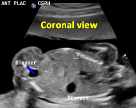
Frontal tangential coronal view two-dimensional ultrasonography in  assessment of fetal face [mouth and nose] in comparison with  four-dimensional ultrasonography | Egyptian Journal of Radiology and  Nuclear Medicine | Full Text
Frontal tangential coronal view two-dimensional ultrasonography in assessment of fetal face [mouth and nose] in comparison with four-dimensional ultrasonography | Egyptian Journal of Radiology and Nuclear Medicine | Full Text

Scanning Planes and the Normal Appearance of the Fetal Spine

PLOS ONE: Sequential Cranial Ultrasound and Cerebellar Diffusion Weighted  Imaging Contribute to the Early Prognosis of Neurodevelopmental Outcome in  Preterm Infants
PLOS ONE: Sequential Cranial Ultrasound and Cerebellar Diffusion Weighted Imaging Contribute to the Early Prognosis of Neurodevelopmental Outcome in Preterm Infants

Coronal ultrasound anatomy of a normal hip in a newborn baby | The BMJ

Scanning School - FAST — Taming the SRU

Scanning Planes and Scanning Methods | Radiology Key

State-of-the-art neonatal cerebral ultrasound: technique and reporting |  Pediatric Research
State-of-the-art neonatal cerebral ultrasound: technique and reporting | Pediatric Research

Right Intercostal Oblique Ultrasound View

11 Neurology ideas | neurology, hemorrhage, ultrasound

References in 3D ultrasound assessment of endometrial junctional zone  anatomy as a predictor of the outcome of ICSI cycles - European Journal of  Obstetrics and Gynecology and Reproductive Biology
References in 3D ultrasound assessment of endometrial junctional zone anatomy as a predictor of the outcome of ICSI cycles - European Journal of Obstetrics and Gynecology and Reproductive Biology

Three‐Dimensional Coronal Plane of the Uterus - Timor‐Tritsch - 2021 -  Journal of Ultrasound in Medicine - Wiley Online Library
Three‐Dimensional Coronal Plane of the Uterus - Timor‐Tritsch - 2021 - Journal of Ultrasound in Medicine - Wiley Online Library

Different planes of fetal ultrasound (US) image samples for training (left  upper is axial plane, right upper is coronal plane, left bottom is sagittal  plane, and right bottom is non-fetal facial standard
Different planes of fetal ultrasound (US) image samples for training (left upper is axial plane, right upper is coronal plane, left bottom is sagittal plane, and right bottom is non-fetal facial standard
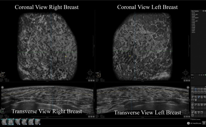
The value of coronal view as a stand-alone assessment in women undergoing  automated breast ultrasound | SpringerLink
The value of coronal view as a stand-alone assessment in women undergoing automated breast ultrasound | SpringerLink

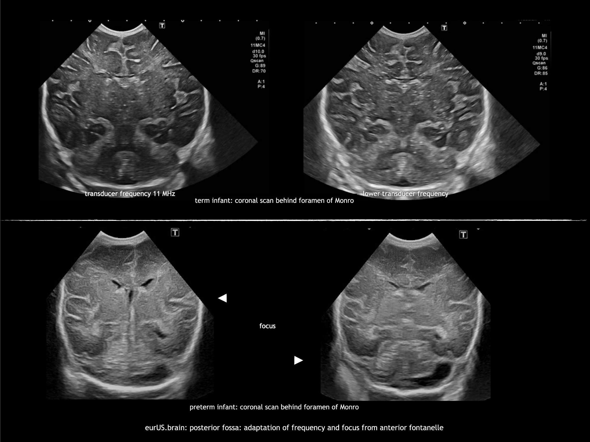
A New Ultrasound Marker for Bedside Monitoring of Preterm Brain Growth |  American Journal of Neuroradiology
A New Ultrasound Marker for Bedside Monitoring of Preterm Brain Growth | American Journal of Neuroradiology

Ultrasound images of the anal canal with bag in place. From the... |  Download Scientific Diagram
Ultrasound images of the anal canal with bag in place. From the... | Download Scientific Diagram

Axial, sagittal, and coronal view of 3D ultrasound imaging. | Download  Scientific Diagram
Axial, sagittal, and coronal view of 3D ultrasound imaging. | Download Scientific Diagram